×
模态框(Modal)标题
在这里添加一些文本
Close
Close
Submit
Cancel
Confirm
×
模态框(Modal)标题
×
Toggle navigation
更多内容请点击
Home
Editorial Department
About Journal
Editorial Board
Instruction
Journal Online
Just Accepted
Current Issue
Archive
Most Read
Most Download
Most Cited
E-mail Alert
RSS
Subscription
Contact Us
中文
Figure/Table detail
The application value of AI-assisted compressed sensing in knee joint MRI
NIE Hongyan, ZHAO Yanjie, XING Wen, CAI Wei
Journal of Jinan University Natural Science & Medicine Edition
, 2024, 45(
6
): 667-673. DOI:
10.11778/j.jdxb.20240021
分组
冠状位T
1
冠状位PD
冠状位T
2
矢状位PD
矢状位T
2
轴位T
2
合计
ACS组
40
51
34
48
38
34
245
对照组
69
71
66
98
73
84
461
Table 2
Comparison of MRI acquisition time of knee joints between ACS group and control group (
t
/s)
Other figure/table from this article
Table 1
Comparison of scanning parameters of knee joint Coronal PD sequence between ACS group and control group
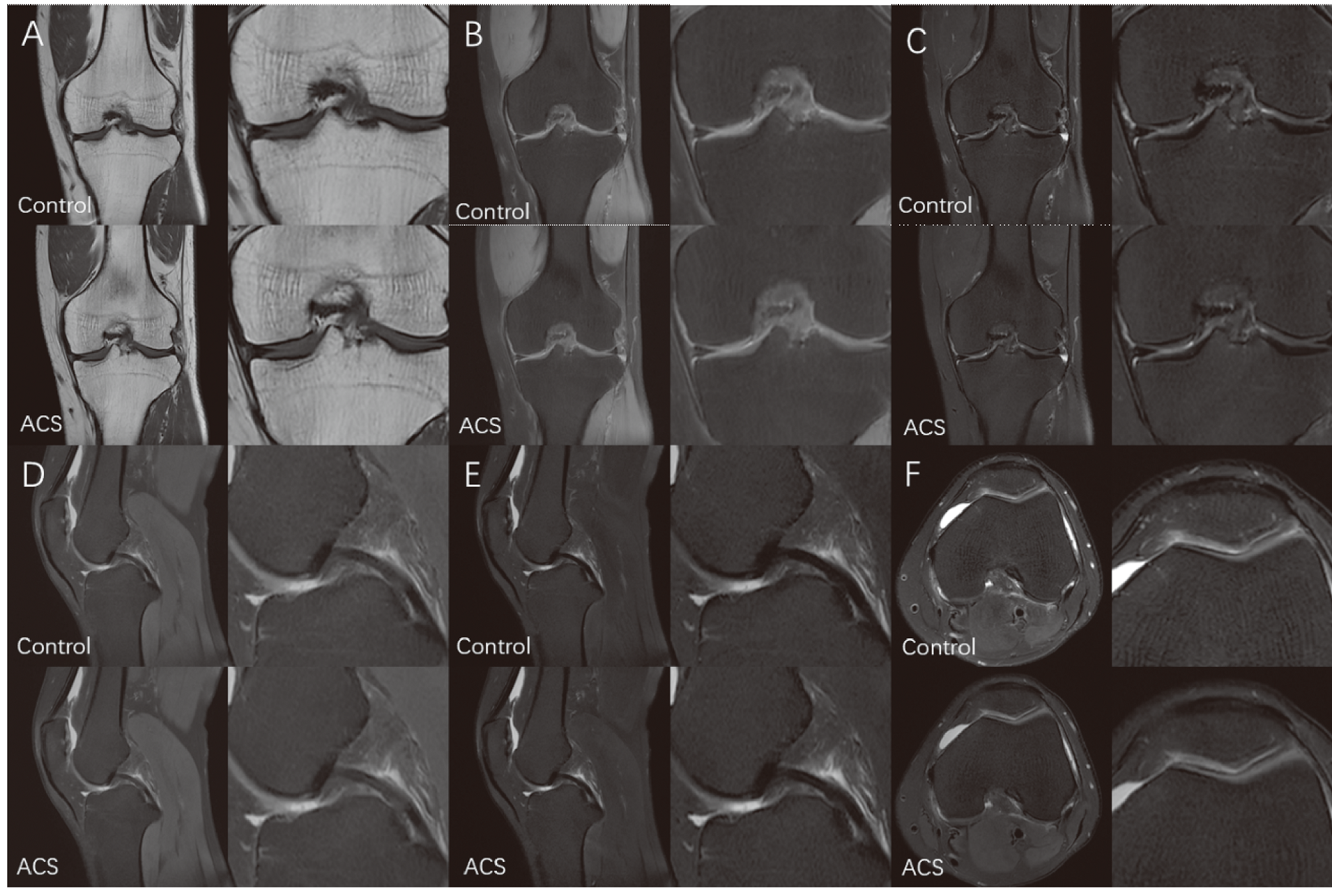
Figure 1
Comparison of knee joint MRI between the ACS group and the control group
A: Coronal T
1
; B:Coronal PD; C:Coronal T
2
; D:Sagittal PD; E:Sagittal T
2
; F:Axial T
2
.
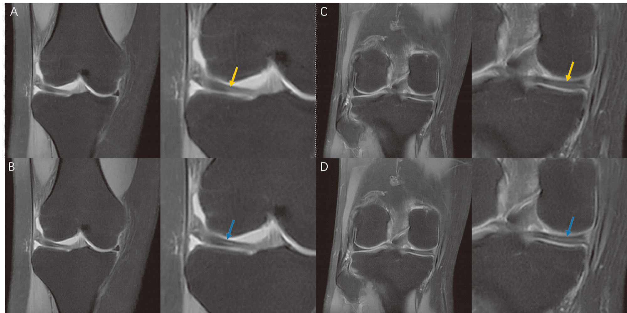
Figure 2
Comparison of MRI images of meniscus injury between ACS group and control group
A、B: A 25 year old female with right lateral discoid meniscus and injury; C、D: A 41 year old male with injury to the posterior horn of the medial meniscus of the right knee; A、C: The PD images of the control group; C、D: The PD images of the ACS group.
Table 3
Consistency comparison of subjective scoring results of two physicians
M
(
P
25
,
P
75
)
Table 4
Comparison of objective evaluation results of image quality between the ACS and control group
M
(
P
25
,
P
75
)


 粤ICP备12087612号