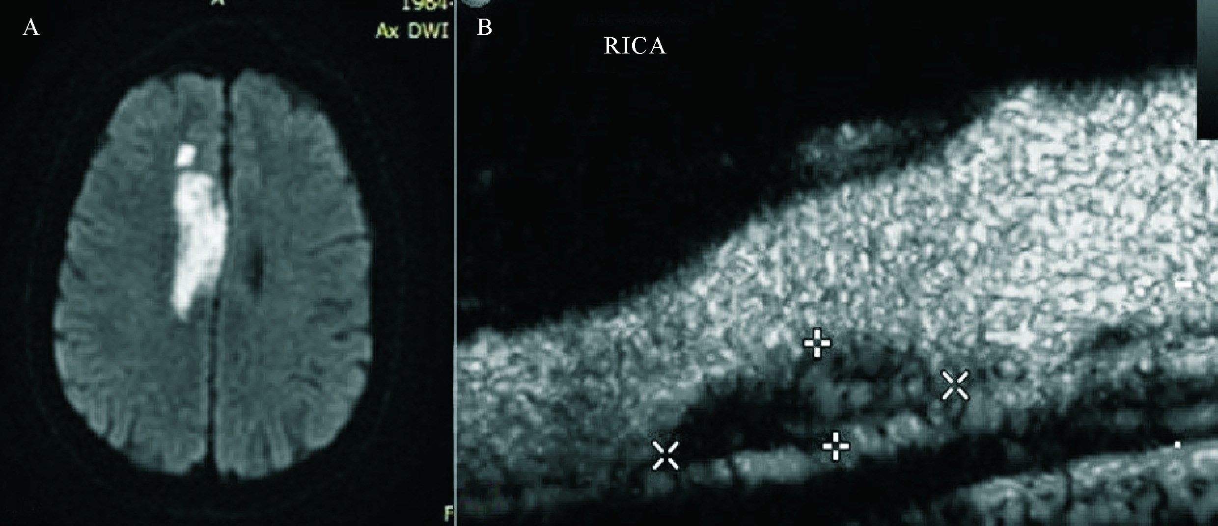
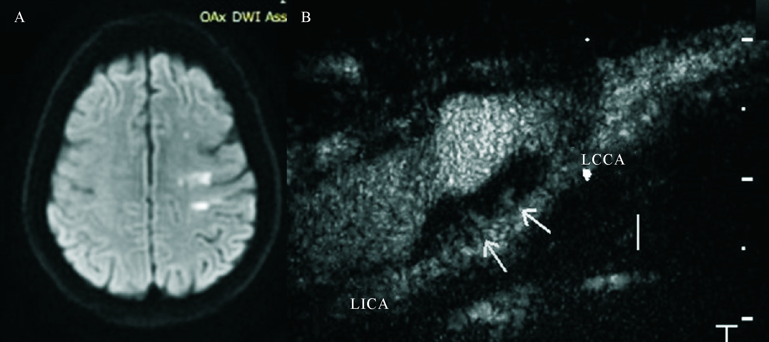
Figure 1 The image of MRI and contrast-enhanced ultrasound of the patient with acute small area cerebral infarction A: MRI of the head of the patient with acute small area cerebral infarction in the left temporo occipital lobe; B: Contrast-enhanced ultrasound of the plaque at the patient’s left internal carotid artery, showed moderate amount of microbubbles (neovascularization is classified as Grade Ⅱ)
Other figure/table from this article


 粤ICP备12087612号